Most often, when a person hears the word "pixel", he immediately thinks about digital cameras, smartphone modules, and other equipment. Naturally, the question arises - how many megapixels are in the eye, because the structure of the eye has something in common with the device of an ordinary camera.
What is a pixel?
The term "pixel" itself began to spread from the moment the number arose. It stands for “picture element,” that is, an image element. A pixel is a point that forms a single picture with other points. A single frame, made in digital format, can contain millions of pixel points.

Each pixel is 5 information elements. Two of them are vertical and horizontal coordinates. The rest are needed to determine the brightness of red, blue and green tones. Together, the elements enable the reader to make the right choice in determining the hue of the point and its subsequent placement.
Interesting fact: A million pixels creates a megapixel, a term that is used more often. In megapixels, the sizes of photos or scanned images are mainly measured.
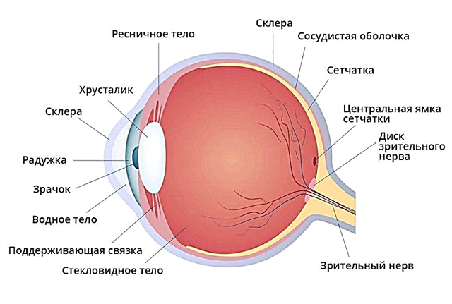
Eye structure
The purpose of the eye is the transmission of the image to the optic nerve. It consists of many components, each of which is very important.
The cornea is a transparent membrane where there are no blood vessels. Despite this, it has a refractive power and is a necessary element of "optics".On the border with it is the outer shell - the sclera.
Between the iris and the cornea there is a space called the anterior chamber. It contains intraocular fluid.
The iris has a colored rounded shape and a hole inside. The iris is the muscles that perform the constriction and expansion of the pupil. Function - regulation of light flow, just like in the camera device. The hole in it is the pupil. The more light, the less the pupil.
The lens is a kind of lens, characterized by transparency and elasticity. Changes shape by focusing on specific objects. The lens allows you to see objects that are nearby or at a distance.

The retina is formed by photoreceptors, as well as nerve endings. They are characterized by special sensitivity. There are two types of receptors: cones and rods. They serve to transform photons into electrical energy of the nervous system, that is, a complex photochemical reaction occurs.
The sclera is a membrane on the outside that passes into the cornea. Muscles are attached to it, with the help of which the eye moves.
Behind the sclera is expelled by the choroid and borders the retina. The membrane supplies blood to the entire structure of the eyeball. Nerves send signals to the brain, and the person sees the image.
Interesting fact: when the retina gets sick, the membrane also suffers from an inflammatory process. However, it lacks nerve endings and pain that would indicate various difficulties.
The number of megapixels in the eye
There are no full-fledged pixels (cells) in the retina matrix of the eyeball. However, there are so-called subpixels that differ in sensitivity and have an uneven arrangement.
Photoreceptor subpixels are divided into cones and rods. The latter perceive only the blue spectral part, and cones - green-yellow, yellow-red and violet-blue. It is impossible to calculate the number of megapixels, since there is no digital matrix in our eye.

In the classical sense, our eyes do not have pixels, since a pixel is a cell. There are no such cells in the retina. The pixels here are photoreceptor cells, which a total of about 126 million.


